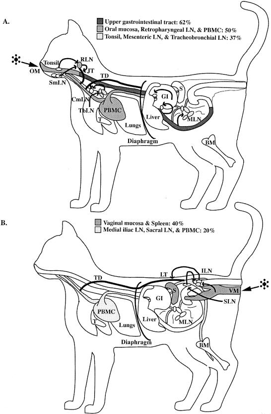
FIG. 2.
(A) Illustration demonstrating target tissue infection frequency between 1 and 3 days after oral-nasal FIV exposure as determined by DNA PCR and RNA ISH. FIV was most frequently identified in tissues of the upper gastrointestinal tract (GI) (62% of the animals). The next most frequently observed FIV-infected tissues were oral mucosa (OM), retropharyngeal LN (RLN), and PBMC (50% of the animals). (B) Illustration demonstrating target tissue infection frequency between 1 and 3 days after vaginal FIV exposure as determined by DNA PCR and RNA ISH. FIV was most frequently identified in tissues of the vaginal mucosa (VM) and spleen (S) (40% of the animals). The next most frequently observed FIV-infected tissues were medial iliac LN (ILN), sacral LN (SLN), and PBMC (20% of the animals). Arrows (both panels), flow of lymphatic drainage from the mucosal tissues to regional lymphoid tissues to lymphatic trunks and/or ducts, which ultimately empty into the venous circulation (PBMC). BM, bone marrow; CmLN, cranial mediastinal LN; JT, jugular trunk; LT, lumbar trunk; SmLN, submandibular LN; T, thymus; TbLN, tracheobronchial LN; TD, thoracic duct.

