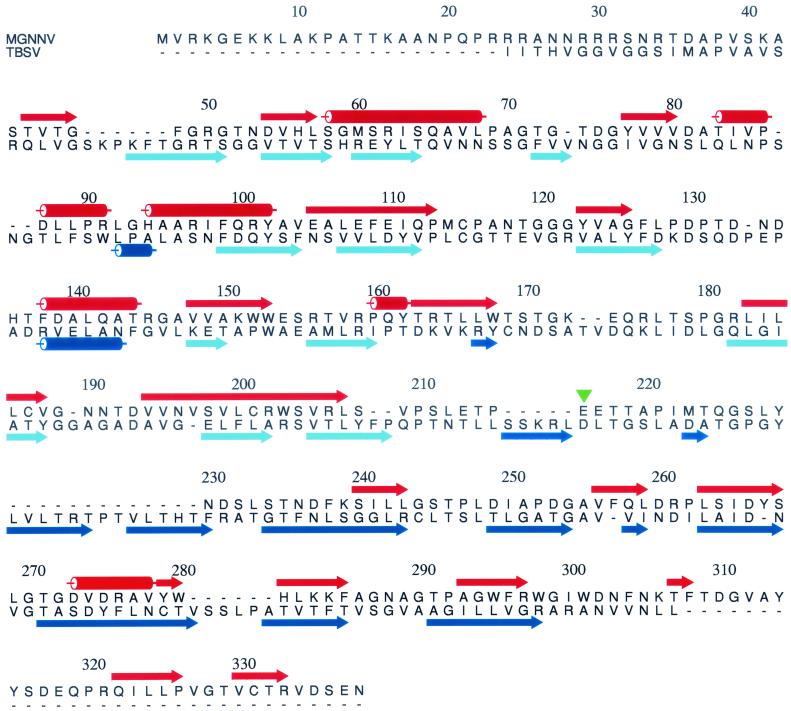
FIG. 6.
Alignment of MGNNV and TBSV coat protein sequences by 3D-PSSM. Although there is no significant sequence identity between the coat proteins of the two viruses, the pattern of secondary structures of MGNNV is rich in β-elements and matches that of TBSV to a reasonable extent. The secondary structures of MGNNV (red) are predicted by 3D-PSSM, and those of TBSV (blue and light blue) are assigned based on the crystal structure. The α-helices and β-strands are shown as rods and arrows, respectively. In TBSV, the β-strands forming the N-terminal β-sandwich domain are colored light blue. The green triangle near residue 216 indicates the boundary between the putative two domains of MGNNV.

