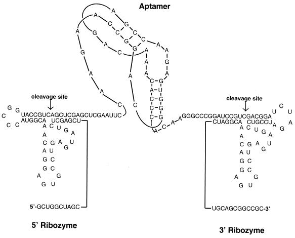
FIG. 1.
Expression of anti-HIV-1 RT aptamer pseudoknots with minimal flanking sequences. A schematic diagram of a representative anti-RT aptamer, 70.28, flanked by self-cleaving ribozymes is presented. The aptamer is represented as a pseudoknot to reflect the secondary structure proposed by Burke et al. (5). The ribozyme sequences are positioned to cleave the required sites according to Benedict et al. (2).

