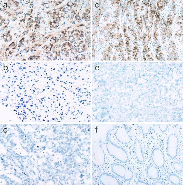
FIG. 3.
IL-1β ISH of the GCs associated or unassociated with EBV. IL-1β ISH was applied to the formalin-fixed and paraffin-embedded tissue sections of the tumor strains and the surgically resected carcinomas. With an IL-1β antisense probe, a positive signal of IL-1β mRNA was observed in the cytoplasm of the KT tumor cells (a), whereas no signal was observed in KN-91 (c). There was no signal in the KT tumor cells when the ISH probe was replaced with the sense probe (b). Similarly, a positive signal was specifically observed in the cytoplasm of the surgically resected carcinoma, which is associated with EBV (d). On the other hand, there was no signal in the surgically resected carcinoma, which was EBV negative (e). The normal gastric mucosa was negative for IL-1β (f). Magnification, ×66.

