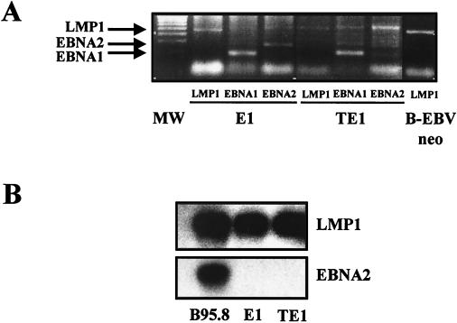
FIG. 3.
E1 and TE1 expressed the EBV genome. (A) Detection of the EBV genome in infected monocytes. The presence of viral DNA in Hirt's extracts was evaluated by PCR amplification of three EBV latent genes: LMP-1, EBNA-1, and EBNA-2. The corresponding PCR products (see primers in Table 2) were visualized by ethidium bromide staining on a 1.8% agarose gel. An LCL was used as a positive control for detection of the viral gene. Lane MW, 100-bp ladder. (B) Detection of viral RNA in E1 and TE1 by RT-PCR and Northern blotting experiments using primers and probes specific for a selected set of viral genes (see primers in Table 2). B95.8 cells were used as a positive control.

