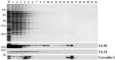
FIG. 8.
Association of UL56 protein with lipid rafts. Cells were solubilized in 1% Triton X-100 and fractionated on a discontinuous sucrose gradient, as described in Materials and Methods. Equal volumes of the recovered fractions were separated by SDS-PAGE and analyzed by silver staining (upper panel) or by Western blotting with anti-UL56 serum, anti-UL34 serum, or anti-caveolin 1 monoclonal antibody. Positions of molecular mass markers (lane P, in kilodaltons) are indicated on the left.

