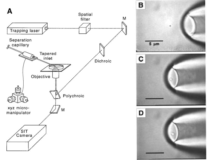
Figure 2.

(A) Schematic of optical trap instrumentation. (Note: dichroic and polychroic mirrors were incorporated into this setup because the instrumentation is sometimes used for simultaneous fluorescence microscopy.25) (B) Video image of trapped single vesicle. (C) Image of a single vesicle at the tapered capillary inlet. (D) Image of single vesicle entering the capillary inlet.
