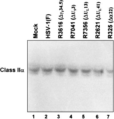
FIG. 5.
Phosphorimage of electrophoretically separated cell lysates reacted with antibody recognizing MHC class IIα chains. Replicate 25-cm2 flask cultures of His16 cells were mock infected or exposed to 10 PFU of the indicated viruses per cell, harvested 12 h after infection, and solubilized. Equivalent amounts of protein from each lysate were electrophoretically separated in a denaturing gel, electrically transferred to a nitrocellulose sheet, and reacted with antibody DA6.147.

