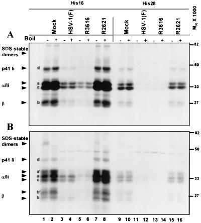
FIG. 7.
Autoradiographic image of electrophoretically separated MHC class II complexes immunoprecipitated with antibody DA6.146 from mock- or virus-infected cell lysates. Replicate 25-cm2 flask cultures of His16 and His28 cells were mock infected or exposed to 10 PFU of the HSV-1(F), Δγ134.5, or ΔUL41 viruses per cell. To radiolabel proteins, cells were incubated in the presence of [35S]methionine for 2 h after infection. Cultures were washed with unlabeled media, and at 2 h (panel A, no chase period), 4 h (2-h chase period, data not shown), or 8 h (panel B, 6-h chase period) after infection, the cells were harvested and solubilized. MHC class II complexes were immunoprecipitated with DA6.147 antibody, separated in 10% denaturing gels, and visualized by autoradiography. Plus and minus signs indicate whether or not samples were boiled. MHC class IIα and -β chains, SDS-stable dimers, and p33 and p41 invariant chain isoforms are indicated on the left with arrowheads. The positions of the molecular weight markers are indicated on the right.

