FIG. 1.
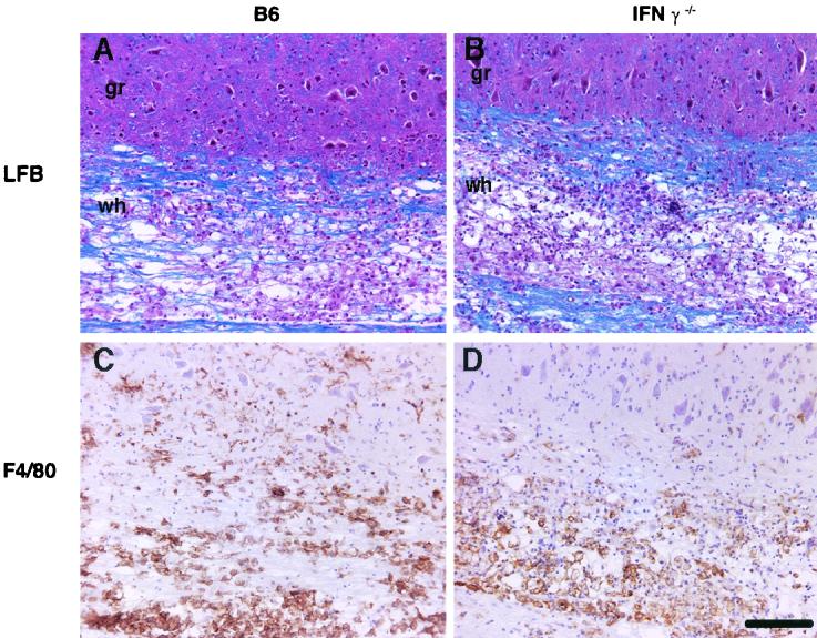
Demyelination and cellular infiltration in recipients of B6 and IFN-γ−/− splenocytes. MHV-infected RAG1−/− mice received CD4 T-cell-enriched splenocytes from immune B6 (A and C) or IFN-γ−/− (B and D) donors at 4 days p.i. Mice were sacrificed at 7 days p.t. (A and B) Representative longitudinal sections of spinal cord were stained with LFB to detect myelin. Although demyelination was detected in both types of recipients, larger amounts were detected in recipients of IFN-γ−/− cells (summarized in Table 1) (C and D) Sections were stained for macrophages/microglia. Equivalent numbers of macrophages/microglia were present in the white (wh) matter of both groups, but the number of these cells in the gray (gr) matter of recipients of B6 cells was significantly greater than in the gray matter of those receiving IFN-γ−/− cells (summarized in Table 3). Scale bar, 200 μm.

