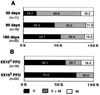
FIG. 5.
Distribution of β-Gal-expressing region in cultured brain slices. (A) Brain slices from mice infected with 5 × 10² PFU 24 h after birth (neonatal infection) maintained for 30, 90, or 180 days. (B) Brain slices from mice infected at 6 weeks of age (young adults) with 5 × 10⁴ or 5 × 10³ PFU and maintained for 180 days. All of the brain slices were cultured for 4 weeks, fixed, and stained with X-Gal. V, X-Gal-positive area only in the ventricular region; M+V, X-Gal-positive area in both regions; M, X-Gal-positive area only in the cerebral marginal region. The ratio was expressed as percentages of numbers of mice with brains of each group. Three brain slices were examined per brain.

