FIG. 7.
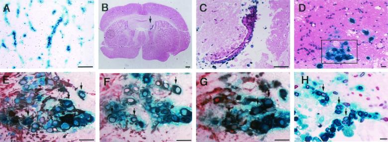
Histochemical and immunohistochemical examination of β-Gal-expressing cells. After X-Gal staining, brain slices were embedded in paraffin and serial sectioned for histological and immunohistological analysis. Vascular distribution of X-Gal-positive cells in 4-week-cultured brain slices from mice infected during the neonatal period and maintained for 90 days (A). X-Gal-positive cells are seen in the ventricular region (arrow) in the 4-week-cultured brain slices from mice infected as young adults and maintained for 180 days (B and C). In order to identify features of the reactivated cells (D [inset]), GFAP (E), X-Gal-positive cells in sections from brain slices cultured for 2 weeks were immunostained with antibodies specific to nestin (F), and Musashi-1 (G). Brain slices were labeled with BrdU for the last 24 h before analysis and stained with the antibody specific to BrdU (H). Size bars: panels A to C, 300 μm; panels D to H, 50 μm. In panels E to H, arrows indicate representative double-stained cells.

