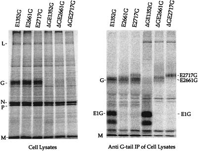
FIG. 2.
Analysis of proteins encoded by VSV recombinants. BHK cells were infected with either VSV-E1352G, VSV-E2661G, VSV-E2717G, VSVΔG-E1352G, VSVΔG-E2661G, or VSVΔG-E2717G at an MOI of 10 and labeled with [35S]methionine. (Left) Cell lysates were analyzed by SDS-10% PAGE. The positions of the five VSV proteins L (241 kDa), G (63 kDa), N (47 kDa), P (30 kDa), and M(27 kDa) are indicated. (Right) BHK cell extracts were immunoprecipitated with a rabbit anti-VSV G tail antibody and analyzed by SDS-10% PAGE. VSV M protein aggregates and appears at substantial levels in all precipitates.

