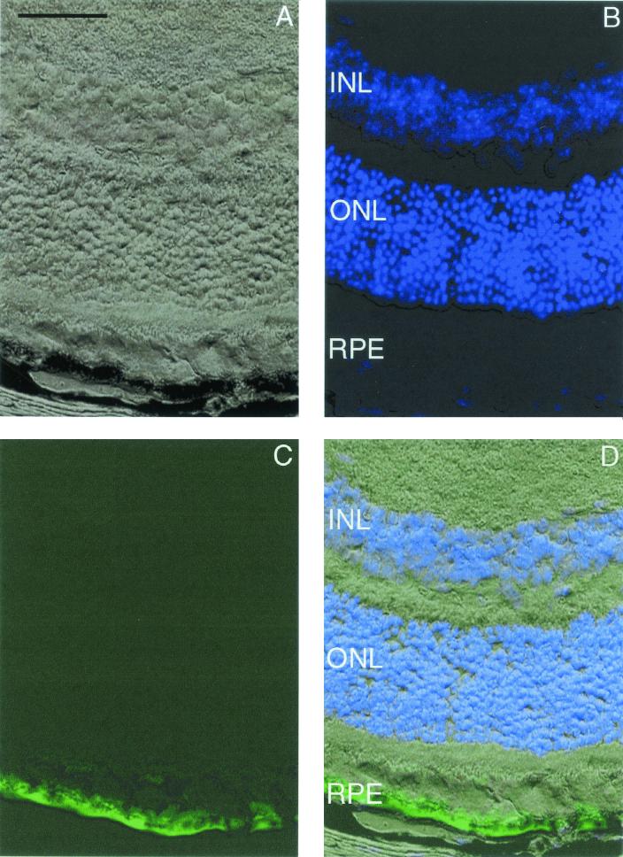
FIG. 11.
Murine retina infected with AAV2/6.GFP 5 weeks after subretinal delivery. (A) Light microscopy of retina using differential interference contrast optics. (B) DAPI-positive nuclei. The area near the RPE has been lightened to allow visualization of DAPI staining. (C) GFP-positive RPE. (D) Superimposed images of panels A to C. Photomicrographs are similar to those taken from retinas harvested 15 weeks after surgery. Bar, 50 μm.

