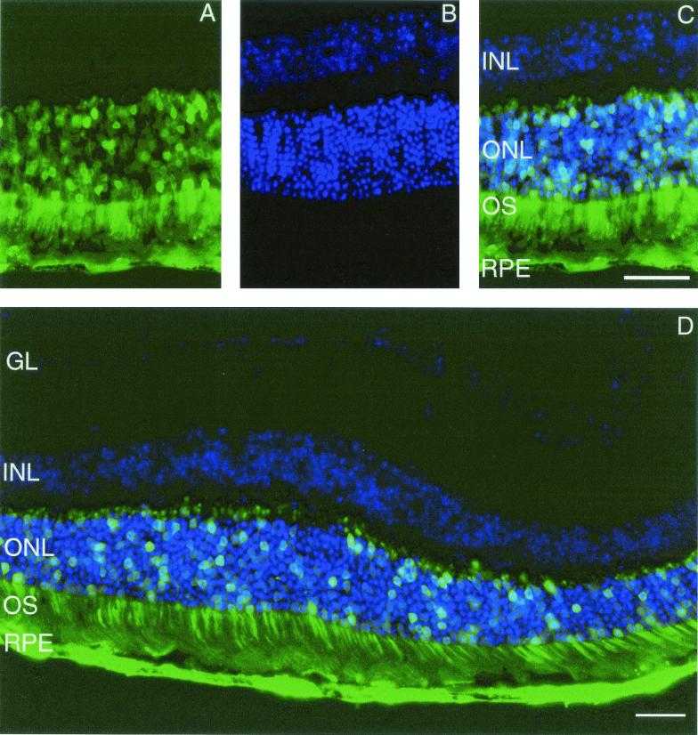
FIG. 6.
Murine retinas infected with AAV5.GFP.Short 5 weeks after subretinal delivery. (A) GFP-positive RPE and photoreceptor cells (ONL and OS [outer segment]). (B) DAPI fluorescence depicting the nuclei within the ONL and INL. (C and D) Superimposed images demonstrating GFP-positive nuclei in the ONL. GL, ganglion layer. The photomicrographs are similar to those taken from retinas infected with AAV5.GFP.Short 15 weeks after injection and from retinas infected with AAV2.GFP.Short and AAV2/5.GFP.Long 5 and 15 weeks after surgery. Bars, 50 μm.

