
Fig. 2. TRAMP/Rosa26-FoxM1b and LADY/Rosa26-FoxM1b double transgenic mice exhibit accelerated growth and development of prostate carcinomas.
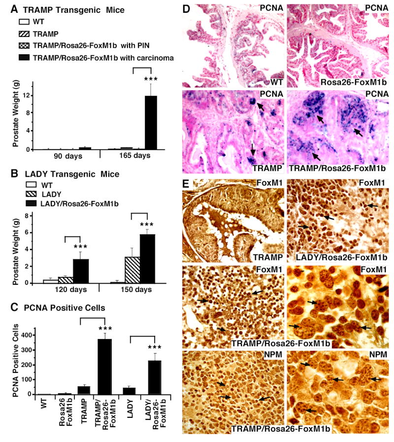
We sacrificed TRAMP and LADY single transgenic (TG) or TRAMP/Rosa26-FoxM1b and LADY/Rosa26-FoxM1b double TG mice at the indicated time points. Prostate glands were collected and weighed from wild type (WT; n=4), Rosa26-FoxM1b (n=7), TRAMP (n=7) and LADY (n=7) single TG mice and TRAMP/Rosa26-FoxM1b (n=12) and LADY/Rosa26-FoxM1b (n=10) double TG mice. Prostate tissue sections were immunochemically stained with antibody specific to either the Proliferation Cell Nuclear Antigen (PCNA) or the FoxM1 protein. (A) Graphically shown are the weights of prostate glands containing carcinomas from TRAMP/Rosa26-FoxM1b double TG mice compared to those of TRAMP TG mice. We determined the mean weight of mouse prostate gland ± SE. Prostate weight is significantly increased in the subset of TRAMP/Rosa26-FoxM1b double TG mice that developed prostate carcinomas compared to dual or single TRAMP TG mice that developed PIN. (B) Statistically significant increase in weight of prostate glands from LADY/Rosa26-FoxM1b double TG mice compared to those of LADY single TG mice. We determined the mean weight of mouse prostate glands ± SE. (C) Elevated number of PCNA positive cells in TRAMP/Rosa26-FoxM1b and LADY/Rosa26-FoxM1b double TG mice. Prostate tumor cells undergoing proliferation were detected with antibody specific to PCNA and prostate tumor sections were then counterstained with nuclear fast red (panel D). We counted the number of PCNA-positive cells in 5 random microscope fields from different mouse prostates to determine the mean number of PCNA positive cells ± SD. The asterisks in panels A, B and C indicate statistically significant increases with P values calculated by Student T Test: *P <0.05, **P ≤ 0.01 and ***P ≤ 0.002. (E) Nucleolar localization of FoxM1 protein in mouse prostate tumors from TRAMP/Rosa26-FoxM1b double TG mice. Prostate tissue sections from TRAMP, TRAMP/Rosa26-FoxM1b and LADY/Rosa26-FoxM1b double TG mice were immunostained with antibody specific to either FoxM1 protein or the nucleolar Nucleophosmin (NPM) protein (49, 50). High magnification of FoxM1 staining in prostate carcinoma cells from TRAMP/Rosa26-FoxM1b double TG mice shows partial nucleolar staining of FoxM1 protein (right middle panel), which is similar to the nucleolar staining pattern of the NPM protein (right lower panel). The nucleolar staining pattern is indicated in these two panels by arrows. Magnification: D and E, 100x; right middle and right lower panels of panel E, 630X.