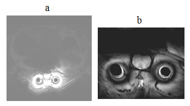
Figure 15.

In vivo axial spin-echo images of spinal cord acquired on day 14 of the coil implantation in (a) larger view of 45 mm × 45 mm and (b) smaller view of 15 mm × 20 mm using the parameters TR/TE = 2500 ms/10 ms, image matrix = 128 × 256, slice thickness = 1 mm and NEX = 2. The image intensity in (a) was windowed and scaled to enhance the background signal.
