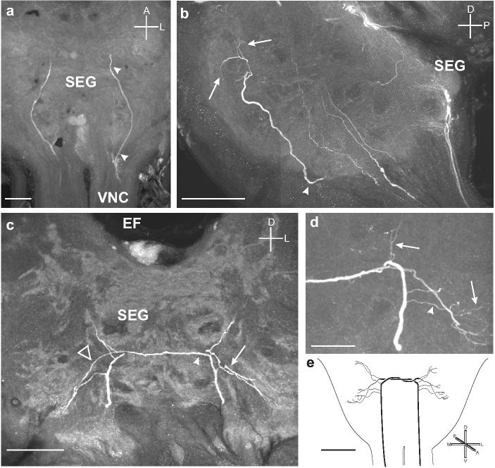
Fig. 10.

a. Horizontal section showing two branches entering the ventral portion of the SEG from each connective of the VNC and extending to the anterior margin of the SEG. b. Sagittal section showing one of the branches (arrowhead) projecting dorsally and producing fine arborizations (arrows) in the dorso-anterior portion of the SEG. c. Frontal section showing each process producing fine arborizations (arrow) and then projecting to the contralateral side of the SEG by following the branch of the other process (arrowhead). The processes then produce finer branches on the contralateral side (open triangle). d. Frontal section through the SEG shows the fine arbors produced by both the ipsilateral (arrowhead) and contralateral (arrows) branches entering the SEG from the VNC. e. Summary diagram of the ascending OAir fibers from the VNC that bilaterally innervate the SEG. Scale bars = 50 μm in a-c, 25 μm in d, and 200 μm in e.
