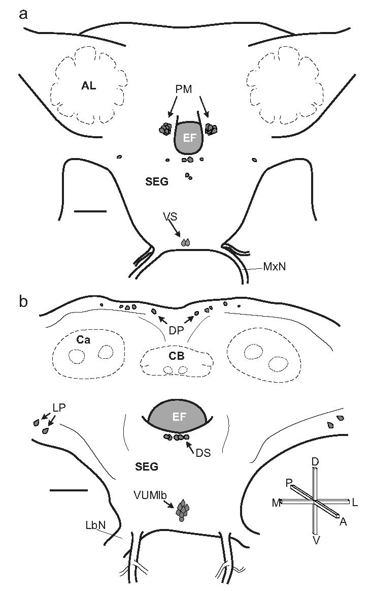
Fig. 2.

Distribution of OAir cell bodies in the brain and SEG of adult M. sexta. a. Frontal view of the anterior portion of the brain (adapted from Homberg et al., 1988). Several of the key cell groups are labeled: two ventral SEG (VS) neurons in the maxillary neuromere, and more dorsally, the bilateral clusters of protocerebral medulla cells (PM) are shown on either side of the esophageal foramen (EF). Also shown are two pairs of dorsal midline cells and two bilateral cell pairs, one of which sits ventro-laterally to the antennal lobes (AL). b. Frontal view of the posterior portion of the brain and SEG (adapted from Homberg and Hildebrand, 1989). Shown in this view is the cluster of mVUM neurons in the labial neuromere, the dorsal SEG (DS) cells, the lateral protocerebral (LP) cells, and the dorsal protocerebral (DP) cells situated above the mushroom body calyces (Ca) and the central body (CB). Scale bars = 200 μm. Crossbars show the orientation of the brain, indicating the posterior (P), anterior, (A), dorsal (D), ventral (V), lateral (L) and medial (M) directions. The same conventions and abbreviations are used for all figures.
