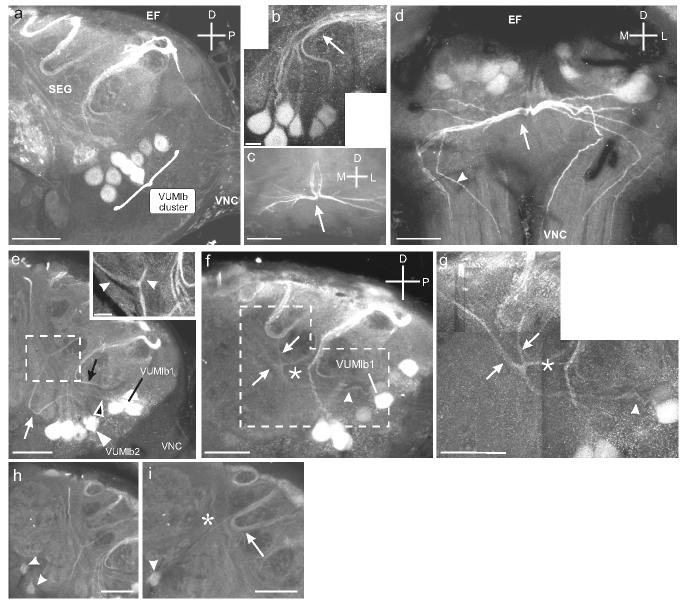
Fig. 3.
Details of the VUMlb cluster and the projections cells in the cluster. a. Sagittal section through the SEG shows the VUMlb (9 cells) situated in the posterior SEG. b. Higher magnification montage of VUMlb neurons projecting dorsally and then posteriorly in a tight bundle (arrow). c. Horizontal section through the dorso-posterior portion of the SEG shows the divergence of the bundle of VUMlb neurites (arrow). d. Frontal section through the SEG shows the diverging bundle and the bilateral projections through the VNC (arrowheads). e. Sagittal section through the SEG showing the VUMlb2 neurites projecting anteriorly before turning dorsally. VUMlb2 (white arrowhead) loops anteriorly before turning dorsally (white arrow). VUMlb1 also projects anteriorly (black arrowhead; see Fig. 4) and then dorsally (black arrow). Inset shows the first branch point of VUMlb2 as it bifurcates below the EF (arrowheads). f. Sagittal section through the SEG shows the neurite of VUMlb1 as it projects anteriorly (arrowhead) and then bifurcates. Inset shows the neurite as it divides, giving rise to bilateral projections traveling dorsally (arrowheads). g. Montage of sagittal section through the SEG shows the two faintly labeled VS neurons (arrowheads). h. Sagittal section through the SEG shows the projection (arrow) of one of the VS neurons (arrowhead) as it extends dorsally and posteriorly, parallel to the neurites of the dorsal SEG (DS; Fig. 7) neurons (asterisk). Scale bars = 50 μm in a, c, d, g-i and 12.5 μm in b, e, f.

