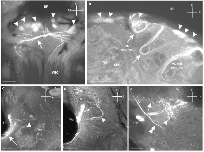
Fig. 5.

a. Frontal section through the SEG shows the dorsal SEG (DS) cells (arrowheads) below the esophageal foramen (EF) and above the branches produced by the VUMlb neurons (arrow) that project down the VNC (Fig. 2d). b. Sagittal section through the SEG shows the primary neurites of the DS cells (arrowheads) as they loop ventrally and then turn back dorsally (arrows). c. Frontal section though the tritocerebrum shows the projections of the DS neurons extending laterally along the ventral side of the EF (arrow) and then branching in the tritocerebrum (arrowhead) below the antennal lobe (AL). d. Frontal section through the tritocerebrum shows some of the branches of the DS neurons (arrowhead) that extend above the PM neurons. e. Sagittal section (detail from Figure 4f) showing the thicker branches of DS neurons as they spread out into both the dorsal (arrow) and ventral (arrowhead) regions of the tritocerebrum. The major ascending processes of VUMlb1 are also clearly visible (double arrowhead). Scale bars = 50 μm.
