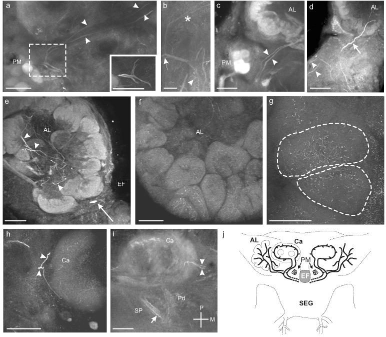
Fig. 6.

a. Sagittal section through the right midbrain showing one branch from each process traveling anteriorly to innervate the AL while the other branch travels dorsally and posteriorly (arrowheads) to innervate the mushroom bodies. Inset shows a higher magnification view of the bifurcation of the two processes before they enter the ALs. b. Sagittal section through the midbrain showing the branches originating from the ventral surface of the esophageal foramen (arrowhead) before bifurcating (arrow) and sending branches anteriorly and posteriorly (asterisk). Scale bar = 12.5 μm. c. Frontal section shows the two processes (arrowheads) entering the ipsilateral antennal lobe (AL). d. Frontal section shows a third process (arrow) entering the AL anterior to the processes from in a-c (arrowheads). e. Frontal section shows numerous branched processes in the coarse neuropil of the AL (arrowheads). A single OAir cell body that sits ventromedial to the AL in the tritocerebrum is also visible (arrow; Fig. 1a). f. Frontal section through the AL reveals fine OAir arborizations in all AL glomeruli. g. Frontal section through the AL shows fine arborizations distributed throughout two glomeruli (dashed outlines). h. Sagittal section through the calyx (Ca) of the ipsilateral mushroom body shows one process branching and entering at the basal ring (arrowheads). i. Oblique horizontal section through the left mushroom body shows the two processes (arrowheads) innervating Ca at the basal ring. OAir processes are also visible in the primary pedunculus (Pd) and the secondary pedunculus (SP) (arrows). Scale bars = 50 μm a, c-i and 12.5 μm in b. j. Summary illustration of the processes innervating the ALs and the MBs.
