
Fig. 7.
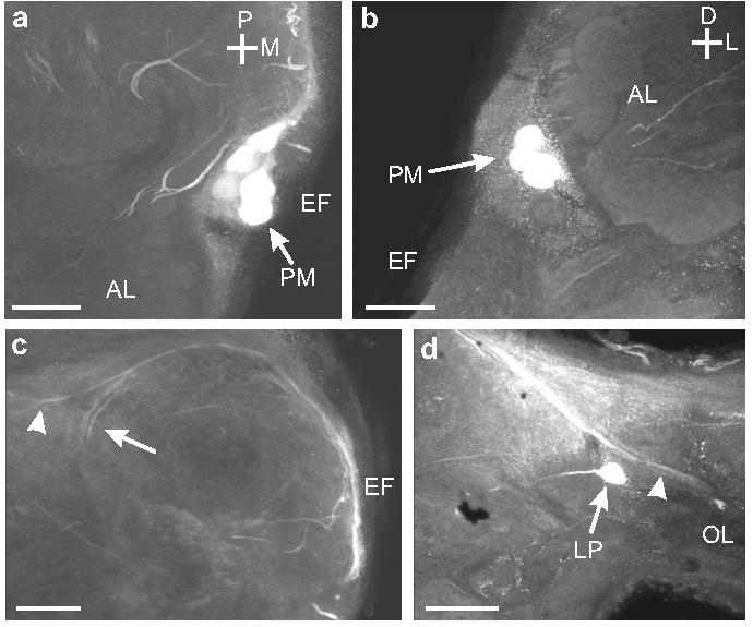
a. Horizontal section through the EF showing the position of the OAir protocerebral medulla (PM) neurons (arrow). This cluster of cells lies posterior and medial to the AL. b. Frontal section through the anterior portion of the brain showing the position of the PM cell bodies (arrows) relative to the EF. c. Horizontal section through the EF shows the OAir projections of the PM neurons into the posterior portion of the brain, then laterally toward the optic lobe (arrowhead), or turning anteriorly in the protocerebrum (arrow). d. Processes (arrowhead) from PM neurons entering the optic lobe (OL). A single lateral protocerebral (LP) neuron is also visible (arrow). Scale bars = 50 μm.