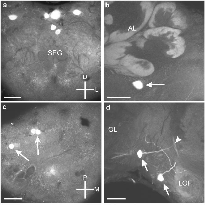
Fig. 9.

a. Frontal section shows the six OAir cell bodies in the dorso-anterior portion of the SEG (see Fig. 1a). b. Frontal section through the tritocerebrum shows the single cell body that sits ventral to the AL (arrow). c. Horizontal section through the protocerebrum shows the three OA-ir cell bodies (arrows) in the dorso-posterior portion of the protocerebrum. d. Frontal section through the tritocerebrum and optic lobe of the left side of the brain shows the two OAir cell bodies (arrows) located dorso-laterally to the lateral optic foci (LOF) near the optic lobes (OL). One of the cells gives rise to a neurite that bifurcates above the LOF (arrowhead). Scale bars = 50 μm.
