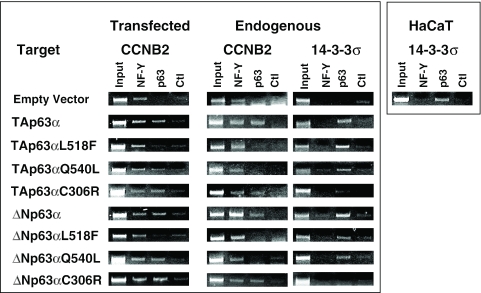
Figure 6.
In vivo recruitment of p63 AEC and EEC mutants to Cyclin B2. ChIP analysis of NF-Y and p63 in SaoS2 cells transfected with ΔN and TA isoforms of p63-wt, L518F, Q540L and C306R- and the control empty vector. Together with the p63 expressing vector, we cotransfected the Cyclin B2 Luciferase construct. The PCRs on the transfected templates are performed with a 5′ primer for Cyclin B2 and a 3′ primer specific for the Luciferase reporter (left panels). The PCRs on the endogenous Cyclin B2 (middle panels) and 14-3-3σ genomic loci (right panels) were performed with the specific oligonucleotides indicated in Materials and Methods on the same immunoprecipitated DNAs. In the right panel, we performed ChIPs with HaCaT chromatin as in Figure 2, to ascertain the positivity of the 14-3-3σ promoter for p63.

