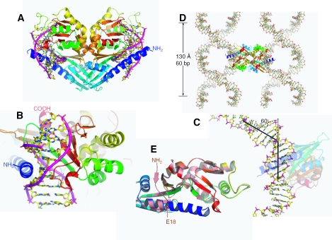
Figure 1.
HinP1I–DNA–Ca2+ structure. (A) The back-to-back HinP1I dimer bound with two DNA duplexes. The HinP1I monomer is colored using a rainbow spectrum with blue for the N-terminus and red for the C-terminus. (B) A HinP1I monomer bound with one DNA duplex, with the N-terminal helix in the minor groove and the central β sheet in the major groove. (C) The DNA is kinked ∼60° where it is bound by the HinP1I enzyme. (D) The 10 bp DNA self-assembles to generate a continuous superhelix. The back-to-back HinP1I dimer links two superhelices together. (E) Structural superimposition of DNA-bound HinP1I (in a rainbow spectrum) with that of free HinP1I (in salmon; PDB 1YNM). The N-terminal residues (amino acids 1–17, colored in blue) become part of a long helix in the DNA-bound structure.

