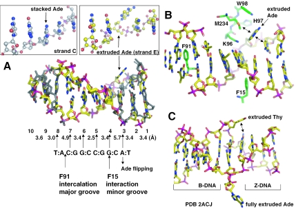
Figure 3.
Base flipping outside of the recognition sequence. (A) Superimposition of the two DNA duplexes, bound with molecule A (colored in grey) or molecule B (colored with yellow for carbon atoms, blue for nitrogen atoms, red for oxygen atoms and magenta for phosphate atoms). Base pairs are numbered from 1 to 10, as shown in Figure 2A. The distances between the stacked bases on strand F are indicated. For comparison, the flipped Ade, A(3) of strand E, and its corresponding A(3) of strand C are enlarged and boxed. (B) The flipped Ade, A(3) of strand E, lies against the HinP1I protein surface of molecule B. The double arrows indicate van der Waals contacts. (C) A view of DNA structure containing a junction between Z-DNA and B-DNA [PDB 2ACJ (17)]. The two bases of a A:T base pair at the junction are extruded. Note the van der Waals interaction (double arrow) between the extruded Thy and an intrahelical base.

