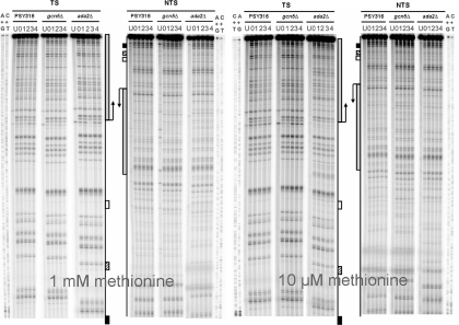
Figure 4.
Representative autoradiographs at nucleotide resolution showing the repair of CPDs in the TS and NTS of the MspI fragment of MET16 in wild type, gcn5Δ and ada2Δ cells grown in 1 mM (repressing) and 10 µM methionine conditions (derepressing). Sanger A+G and T+C sequencing ladders are included to determine the position of the CPDs induced. U, untreated cells; 0, cells treated with UV light no repair; (1–4), UV-treated cells allowed to repair the damage for 1–4 h. The intense top band corresponds to the undamaged MspI fragment of MET16, the bands below represent DNA fragments with a CPD lesion that was cut by the CPD-specific endonuclease. Cbf1p-binding site (closed box), Gcn4p-binding site (stripped box), TATA-box (open box) and coding region (grey box). Note: differences in band migration at the bottom of the autoradiographs are due to the fact that ada2Δ samples were run in different gels to PSY and gcn5Δ.

