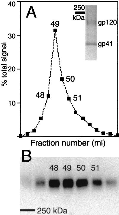
FIG. 1.
Gel filtration analysis of virion-derived HIV-1 Env. (A) Aliquots of gel filtration fractions were subjected to SDS-8% PAGE in the presence of reducing agent (cross-links broken) and sequentially immunoblotted with an HIV-1 gp120-specific MAb, a mouse immunoglobulin G-specific rabbit polyclonal serum, and iodinated protein A. gp120 was quantified by phosphor-screen autoradiography. Blue Dextran 2000 (giving the void volume) had an elution peak in fraction 43. (Inset) Pool of fractions 48 to 51 analyzed by SDS-4 to 20% PAGE in the presence of reducing agent and Coomassie blue staining. (B) Aliquots of gel filtration fractions analyzed by SDS-5% PAGE in the absence of reducing agent (cross-links maintained) and immunoblotted as described above. The bars in panels A (inset) and B indicate the electrophoretic mobility of a 250-kDa marker protein.

