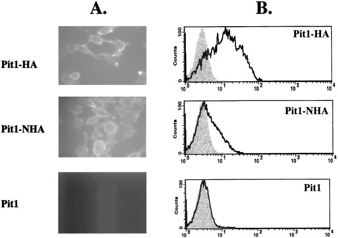
FIG. 6.
. Fluorescence images (A) and flow-cytometric histograms (B) detect the presence of both the N and C termini of the Pit1 receptor on the surfaces of live nonpermeabilized MDTF cells expressing Pit1 receptors bearing either C- or N-terminal HA epitope tags (Pit1-HA and Pit1-NHA, respectively). Pit1 bearing no epitope tag (Pit1) was used as a negative control. Cells were incubated with monoclonal antibody HA.11, washed, and then incubated with a fluorescein-conjugated second antibody. Shaded areas on histograms represent negative-control MDTF-Pit1 cells; areas beneath the boldface lines represent MDTF cells expressing HA-tagged receptors. The x and y axes are as defined for Fig. 2.

