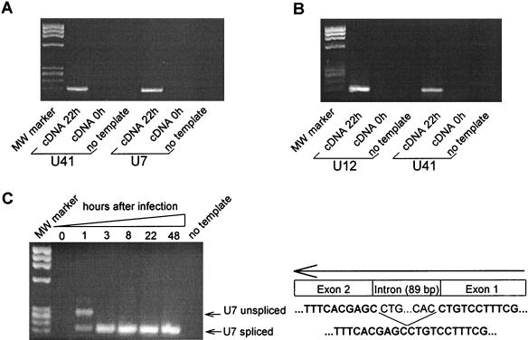
FIG. 1.
Early detected viral transcripts in HHV-6B-infected Molt-3 T cells do not originate from contaminating viral RNA or DNA. (A) Viral RNA is not detected in T cells after a few minutes of HHV-6B infection. Agarose gel electrophoresis of the products from 45 cycles of real-time PCR on cDNA derived from T cells infected by HHV-6B for 22 h (lane 1 and 4) or from T cells briefly mixed with virus supernatant (lane 2 and 5). Lanes 3 and 6, no template control. MW, DNA Molecular Weight Marker IX (Roche). Products in lanes 1, 2, and 3 were amplified with the primer pair for gene U41; products in lanes 4, 5, and 6 were amplified with the primer pair for gene U7. (B) RNA preparations from HHV-6B-infected T cells do not contain contaminating viral DNA. A cDNA reaction mixture not retrotranscribed was amplified for 45 cycles of real-time PCR by using primers for U12 (lanes 1, 2, and 3) and U41 (lanes 4, 5, and 6) and was separated by agarose gel electrophoresis. Lanes 1 and 4, HHV-6B-infected T cells (48 h). Lanes 2 and 5, RNA was purified from T cells briefly mixed with virus supernatant and cDNA reactions performed in the absence of retrotranscription. Lanes 3 and 6, no template control. MW, DNA Molecular Weight Marker IX. (C) Induction of a spliced form of U7 indicates de novo RNA synthesis. Agarose gel electrophoresis of the products from 45 cycles of real-time PCR on cDNA from uninfected (lane 1) and HHV-6B-infected T cells (lanes 2 to 6) is shown. Lane 7, no template control. All products have been amplified with the primer pair for gene U7. MW, DNA Molecular Weight Marker IX. The GenBank accession number for the U7 sequence is shown in Table 2.

