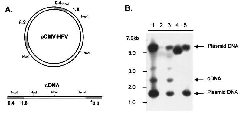
FIG. 5.
Detection of full-length PFV DNA in transfected cells. 293T cells were transfected with CMV-driven viral plasmids or empty (mock) plasmid, and genomic DNA was isolated 4 days posttransfection and digested with NcoI. (A) Diagram of NcoI sites in the viral plasmid used for transfection and predicted sizes of resulting products that hybridize to the probe. The shaded regions of the DNA indicate the locations of the LTRs. The asterisk indicates the size of the fragment corresponding to the unique cDNA fragment. (B) Southern blot of digested DNAs using an LTR-specific radiolabeled probe: lane 1, wild-type HFV; lane 2, mock; lane 3, IN(−); lane 4, PolΔ5; lane 5, RT-V313M. A 1-kb ladder (New England Biolabs) was run alongside the samples for size orientation.

