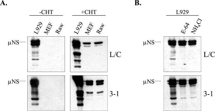
FIG. 6.
Capacity of PI virus isolates L/C and 3-1 to replicate in L929, MEF, and RAW cells. (A) L929, MEF, and RAW cells were infected at an MOI of 5 with PI virus isolates 3-1 and L/C. After adsorption, samples were incubated in media that either did or did not contain CHT at 10 μg/ml (+ CHT and − CHT, respectively). At 15 h p.i., cell lysates were prepared and μNS synthesis was analyzed by immunoblotting as described in the legend to Fig. 1. (B) Control L929 cell infections were performed with the PI viruses at an MOI of 5 in the presence of NH4Cl or E-64, as described in the legend to Fig. 3. Analysis of viral protein expression was performed at 15 h p.i., as described in the legend to Fig. 1.

