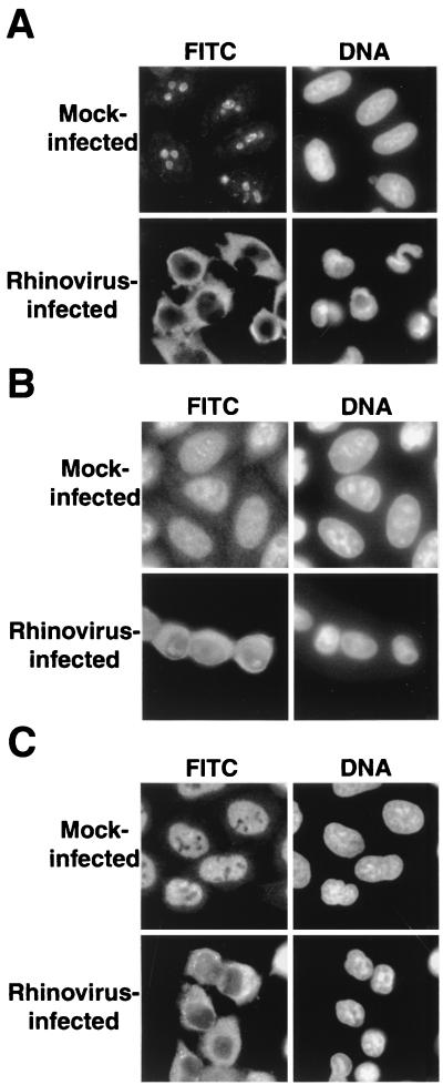
FIG. 1.
Intracellular localization of endogenous cellular proteins in uninfected and rhinovirus-infected cells. (A) Uninfected cells (mock infected) or HeLa cells infected with rhinovirus for 6 h were fixed and stained with an antibody directed against nucleolin. In the FITC panels, cells were examined with an FITC filter to detect the indicated antibodies. In the DNA panels, the same field was examined with a UV filter to visualize Hoechst staining of nuclei. (B) Visualization of La was performed as described for panel A. (C) Visualization of Sam68 was performed as described for panel A.

