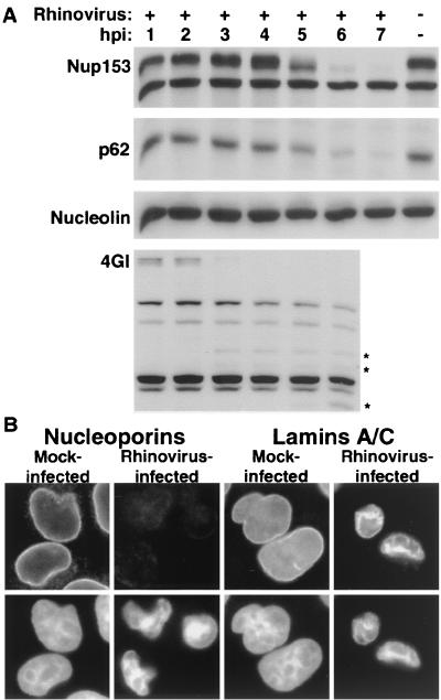
FIG. 6.
Analysis of nuclear pore complex composition in rhinovirus-infected HeLa cells. (A) Fifty micrograms of whole-cell lysates prepared from mock-infected cells or cells that had been infected with rhinovirus for the indicated length of time was analyzed by immunoblotting with monoclonal antibody 414 to detect Nup153 and p62 or MS3 to detect nucleolin. eIF4GI was detected with rabbit polyclonal sera. An asterisk indicates rhinovirus-specific degradation products of eIF4GI. hpi, hours postinfection. (B) Indirect immunofluorescence with monoclonal antibodies 414 and SC-7292 to detect nucleoporins and lamins, respectively. Cells were either uninfected (mock infected) or infected with rhinovirus for 6 h (rhinovirus infected). The top panels show cells examined with an FITC filter, and the bottom panels show the same fields examined with a UV filter to detect Hoechst staining. FITC images for a given antibody were acquired with identical exposure times and adjustments.

