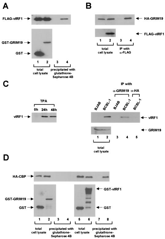
FIG. 3.
Association of vIRF1 and GRIM19 in vivo. (A) A GST expression plasmid (pEBG) or a GST-GRIM19 expression plasmid (pEBG-GRIM19) was cotransfected with FLAG-vIRF1 expression plasmid (pFIN-vIRF1) into 293T cells. Cell extracts were precipitated with glutathione-Sepharose 4B. The resulting precipitates were washed and resolved by SDS-PAGE. GST fusion protein and FLAG-vIRF1 were detected by Western blotting with anti-FLAG (top panel) and anti-GST (bottom panel) antibodies, respectively. Lanes: 1 and 3, GST with FLAG-vIRF1; 2 and 4, GST-GRIM19 with FLAG-vIRF1. (B) An HA-GRIM19 expression plasmid (pcDNA3-HA-GRIM19) was cotransfected with or without the FLAG-vIRF1 expression plasmid (pFIN-vIRF1). Cell extracts were immunoprecipitated with anti-FLAG monoclonal antibody, washed, and resolved by SDS-PAGE. HA-GRIM19 and FLAG-vIRF1 were detected by Western blotting with anti-HA (top panel) and anti-FLAG (bottom panel). Lanes: 1 and 3, HA-GRIM19 alone; 2 and 4, HA-GRIM19 with FLAG-vIRF1. IP, immunoprecipitation. (C) vIRF1 associates with GRIM19 in KSHV-infected BCBL-1 cells. BCBL-1 cells were treated with TPA as previously described (36) and cell extracts were prepared after the indicated number of hours. vIRF1 was detected by Western blotting with anti-vIRF1 polyclonal antibody (left panel). A coimmunoprecipitation assay was performed with BJAB and BCBL-1 cells (2 × 10⁷ cells) after 48 h of TPA induction. Total cell extracts were immunoblotted with anti-vIRF1 and anti-GRIM19, respectively (lanes 1 and 2). Cell extracts were immunoprecipitated with anti-GRIM19 monoclonal antibody (lanes 3 and 4) and anti-HA monoclonal antibody (lane 5). vIRF1 was detected by Western blotting with anti-vIRF1 polyclonal antibody (right top panel). (D) GRIM19 does not interact with CBP. A GST expression plasmid (pEBG), a GST-GRIM19 expression plasmid (pEBG-GRIM19), or a GST-vIRF1 expression plasmid (pEBG-vIRF1) was cotransfected with HA-CBP expression plasmid (pCMV-HA-CBP) into 293T cells. Cell extracts were precipitated with glutathione-Sepharose 4B. The resulting precipitates were washed and resolved by SDS-PAGE. GST fusion proteins and HA-CBP were detected by Western blotting with anti-HA (top panel) and anti-GST (bottom panel) antibodies. Lanes: 1 and 3, GST with HA-CBP; 2 and 4, GST-GRIM19 with HA-CBP; 5 and 7, GST with HA-CBP; 6 and 8, GST-vIRF1 with HA-CBP.

