FIG. 2.
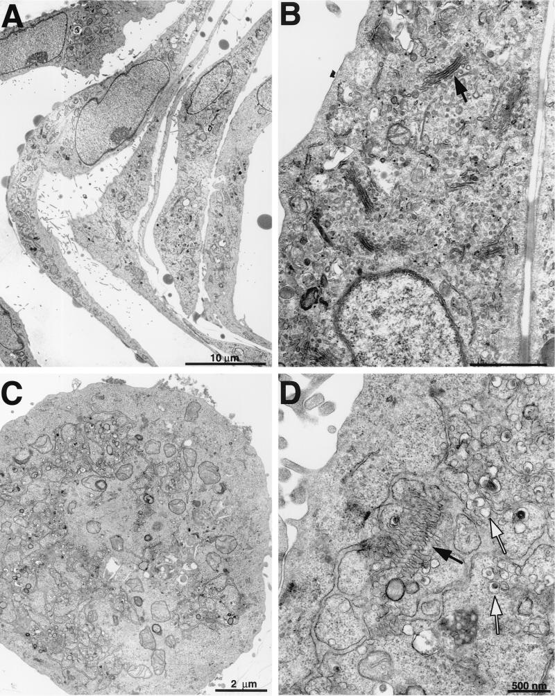
Membrane rearrangements in FCV-infected cells. Transmission electron microscopy of mock- or FCV-infected CRFK cells obtained 6 h postinfection. (A) Mock-infected cells at low magnification showing elongated appearance. (B) Mock-infected cells at higher magnification showing normal intracellular arrangement of membranes (arrow, Golgi apparatus). (C) FCV-infected cells at low magnification showing a rounded appearance. (D) FCV-infected cells at higher magnification showing membrane rearrangements (black arrow, putative Golgi apparatus) and vesicles (white arrows).

