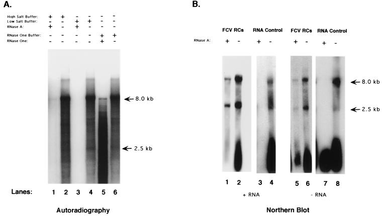
FIG. 4.
(A) 32P-labeled RNA was purified from an FCV-infected RC pellet replication assay and subjected to the following treatments: lane 1, high-salt buffer (2× SSC) with RNase A; lane 2, high-salt buffer only; lane 3, low-salt buffer (0.2× SSC) with RNase A; lane 4, low-salt buffer only; lane 5, RNase One buffer plus RNase One enzyme; lane 6, RNase One buffer only. (B) Northern blot analysis of RNA purified directly from FCV RCs collected at 7 h after infection. Lanes: 1 to 4, RNA probed with an antisense RNA probe that detects positive-sense RNA; 5 to 8, RNA probe with a sense RNA probe that detects negative-sense RNA. RNA in lanes 1 and 5 was purified from RCs that had been treated with RNase A (0.2× SSC) for 1 h. The RNA in lanes 3 and 7 was purified from RCs prior to RNase A treatment.

