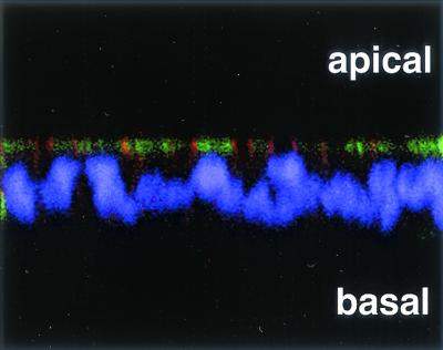
FIG. 3.
DAF is expressed at the apical surface of polarized T84 cells. Polarized T84 monolayers were fixed, permeabilized, and stained for DAF by using the anti-DAF antibody IA10 directly conjugated to FITC (green) and for CAR by using an affinity-purified anti-CAR antibody followed by a secondary antibody conjugated to Cy3 (orange-red). Nuclei were stained with DAPI (purple). Stained cells were examined by confocal microscopy; a representative x-z axis cross section of the monolayer is shown.

