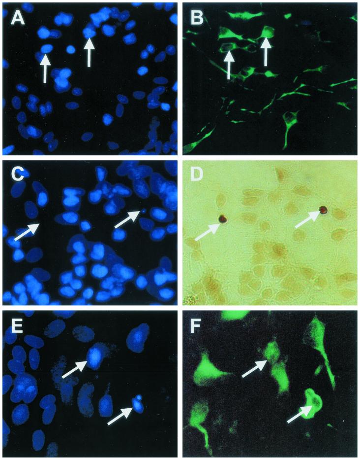
FIG. 2.
Identification of neuronal apoptosis in NT2.N-astrocyte cultures. NT2.N neurons were cultured on a feeder layer of rodent astrocytes for 5 weeks, exposed to HIV-1-infected or noninfected (control) MDM supernatants for 48 h, and then examined by TUNEL assay, Hoechst 33342 staining, or indirect immunofluorescence labeling with an anti-MAP-2 antibody, as described in Materials and Methods. Panels A and B show control cultures colabeled with Hoechst 33342 (A, nuclei) and MAP-2 (B, neurites). Panels C to F represent HIV/MDM-exposed cultures. Panel C shows loss of Hoechst nuclear staining in neurons colabeled by TUNEL (D, arrows). Panel E shows higher magnification of Hoechst-labeled nuclei with morphological features of apoptosis in neurons colabeled with MAP-2 (F). Magnification: panels A to D, ×600; panels E and F, ×1,000.

