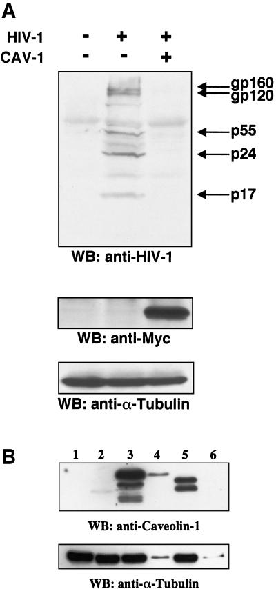
FIG. 5.
(A) Cav-1 inhibits intracellular HIV-1 structural protein accumulation. 293T cells were cotransfected with 1.2 μg of pYU-2 and 0.3 μg of pCMV-myc or pCMV-myc.Cav-1. HIV protein expression was determined 40 h later in cellular lysates by immunoblotting with pooled HIV-1+ human serum. Equal amounts of protein (40 μg) were loaded in each lane. The same membrane was subsequently immunoblotted with an anti-Myc (for Cav-1) and an anti-α-tubulin MAb. HIV-1 p24 in the supernatants of these cells was <1 (mock), 685 ± 49 (pCMV-myc), and 63 ± 16 (pCMV-myc.Cav-1) ng/ml. (B) Cav-1 expression in the transfected 293T cells used in panel A relative to endogenous levels in other cells. The primary antibody is specific for residues 61 to 71 of Cav-1. Lane 1, pCMV-myc-transfected 293T cells (20 μg of protein); lane 2, HeLa cells (20 μg of protein); lanes 3 and 4, pCMV-myc.Cav-1-transfected 293T cells (20 and 0.8 μg of protein, respectively); lanes 5 and 6, primary human VSMCs (20 and 0.8 μg of protein, respectively). Prominent alpha and beta isoforms can be detected in primary human VSMCs and 293T cells, but only the beta isoform was detectable in HeLa cells; the bands in the 293T lanes are slower migrating because of the Myc tag. WB, Western blotting.

