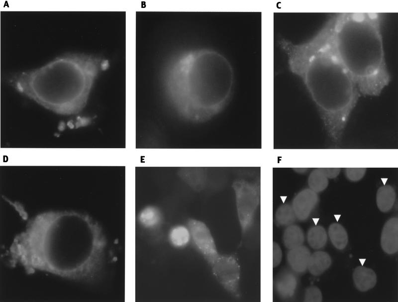
FIG. 7.
Immunofluorescence of Myc-tagged caveolins in 293T cells. (A) Wild type (Cav-11-178); (B) Cav-11-135; (C) Cav-1101-178; (D) Cav-1101-135; (E and F) Cav-11-101. Panel F shows DAPI (nuclear) staining of cells in panel E, and arrowheads indicate nuclei of transfected cells. Of note, most Cav-11-101-transfected cells (80%) showed the nuclear localization found in the two leftmost cells in panel E; this field, in which a lower percentage has nuclear localization, was selected to show representative examples of the remaining cells in which the protein is also seen in the cytoplasm.

