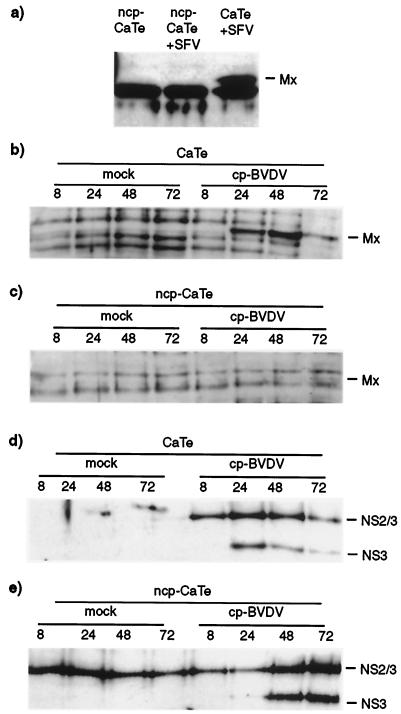
FIG. 3.
ncp BVDV inhibition of virus induction of an IFN-stimulated gene product, MxA. (a) ncp BVDV inhibition of MxA induction by SFV in CaTe cells. Cells were either mock infected or infected with ncp BVDV (1 to 2 PFU/cell) for 48 h. ncp BVDV-infected and mock-infected cultures were subsequently infected with SFV (0.01 PFU/cell) and incubated for 18 h. Cells were harvested in sample buffer and subjected to immunoblot analysis with a rabbit antiserum against human MxA protein. (b through e) Time course of induction of MxA and expression of viral NS3 and NS2/3 by cp BVDV in ncp BVDV-infected CaTe cells. CaTe cells were either mock infected for 48 h (b and d) or infected with ncp BVDV (1 to 2 PFU/cell) for 48 h (c and e). Cells were then either mock treated or infected with cp BVDV (2 to 3 PFU/cell). At the times indicated, cells were harvested in sample buffer and subjected to immunoblotting using either an antiserum against human MxA (b and c) or a bovine antiserum against BVDV proteins (d and e). MxA (a and b) or its expected position (c) is indicated. NS2/3 and NS3 (d and e) are BVDV polypeptides.

