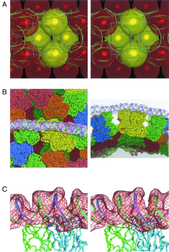
FIG. 2.
Ropes of RNA within BTV core crystals. (A) Stereo figure showing the meshing together of the BTV core particles in the crystals by the long ropes of external dsRNA. The model for the dsRNA is drawn as a green-and-red worm; the core particles are shown to scale as colored yellow and red spheres. (B) Two orthogonal views of the interactions of the parts of one RNA rope with the BTV core. The RNA model and electron density are shown (the density is rendered semitransparent). The VP7 trimers are colored as described in reference 7, with the underlying VP3 molecules shown in slightly darker colors (again, colored as in reference 7). (C) A stereo image of the fit of the dsRNA model, with a portion of the difference electron density map in red, showing the typical disposition of the RNA on the top surface of a VP7 trimer. The strands of the RNA model are shown as green and blue ribbons, and the three polypeptide chains of VP7 are drawn in green, cyan, and blue.

