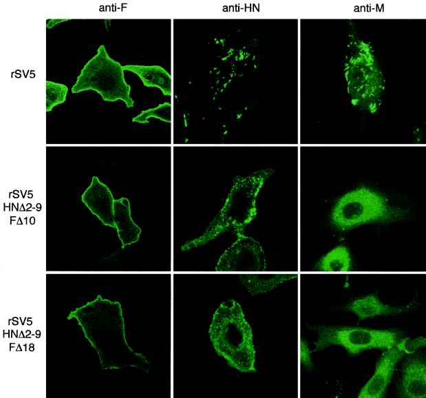
FIG. 8.
Localization of HN, F, and M in cells infected with rSV5 harboring HN and F double cytoplasmic tail truncations. CV-1 cells on glass coverslips were infected with rSV5, rSV5 HNΔ2-9 FΔ10, and rSV5 HNΔ2-9 FΔ18. At 16 h p.i., the surface localization of HN and F proteins was detected by binding HN and F specific MAbs (HN1b and F1a) to intact cells. Intracellular localization of M protein was detected by binding of M-h MAb to saponin-permeabilized cells. The cells were then stained with a FITC-conjugated goat anti-mouse secondary antibody. Fluorescence was detected by using a Zeiss LSM 410 confocal microscope.

