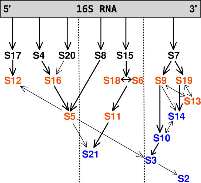
Figure 1. The 30S Subunit In Vitro Experimental Assembly Map for E. coli .
The primary, secondary and tertiary binding proteins are shown in black, orange, and blue, respectively. Arrows indicate facilitating effect of binding of one protein on another. Adapted from the review of Culver [6] based on the work of the Nomura Laboratory [4,5].

