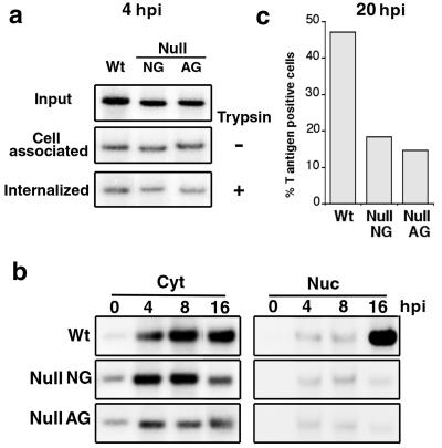
FIG. 3.
Detection of viral DNA in subcellular fractions. (a) Cells infected with the VLP lysate were harvested at 4 hpi either by scraping off (trypsin −, cell associated) or by trypsin treatment (trypsin +, internalized). Low-molecular-weight DNA was extracted and screened for viral DNA by Southern blotting. “Input” represents 1/50 of the viral DNA in total VLP lysate. Each lane of the cell-associated or internalized DNA panel was loaded with 1/20 of the DNA samples. (b) VLP-infected cells were harvested and separated into subcellular fractions at 0, 4, 8, or 16 hpi. Viral DNA in each fraction was detected as for panel a. One twenty-fourth of the cytoplasmic (Cyt) or 1/8 of the nuclear (Nuc) fraction was used for the analysis. (c) VLP-infected cells were fixed at 20 hpi and examined immunocytochemically for T-positive cells. Percent T-positive cells indicates the proportion of cells with visible T antigen in the 2,000 examined cells. Wt, wild type.

