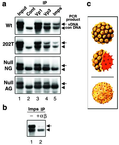
FIG. 6.
Coimmunoprecipitation of viral DNA by antibodies to capsids or cellular importins. (a) The cytoplasmic fraction from respective VLP-infected cells was immunoprecipitated with anti-mouse IgG (Cont), anti-Vp1 (Vp1), anti-Vp3 (Vp3), or a mixture of anti-importin α and anti-importin β antibodies (Imps). The respective immunocomplexes were mixed with a standard DNA, and their DNA was extracted and subjected to semiquantitative PCR for viral DNA. The amplification of the NO-SV40 genome generates the 2.2-kbp fragment (arrow, vDNA) and the standard DNA, the 1.7-kbp PCR fragment (arrowhead, con DNA). In “Input” (lane 1), 10 μl of each cytoplasmic fraction was mixed with the standard DNA, and the extracted DNAs were subjected to PCR. The total viral DNA in the respective cytoplasmic fractions was estimated as 20, 20, 15, or 10 pg for each 50 μl of the wild-type (Wt), KTKRK (202T), Null NG, or Null AG particle-infected cytoplasm, respectively. (b) The wild-type-infected cytoplasmic fraction was immunoreacted with anti-importin in the absence (−) or presence (+αβ) of recombinant GST-importins α and β to which a fivefold excess over the concentration of endogenous importins was added. Viral DNA in each immunoprecipitate was detected as for panel a. (c) Illustrations of an SV40 virion and an internalized particle. Shown are an intact SV40 particle with some of the 72 Vp1 pentamers (top panel, yellow) and a cross section revealing the internal core composed of minor capsid proteins Vp2 and Vp3 and a minichromosome (middle panel, red). A possible alteration of the internalized particle exposing a portion of internal minor capsid proteins on the surface is also shown (bottom panel).

