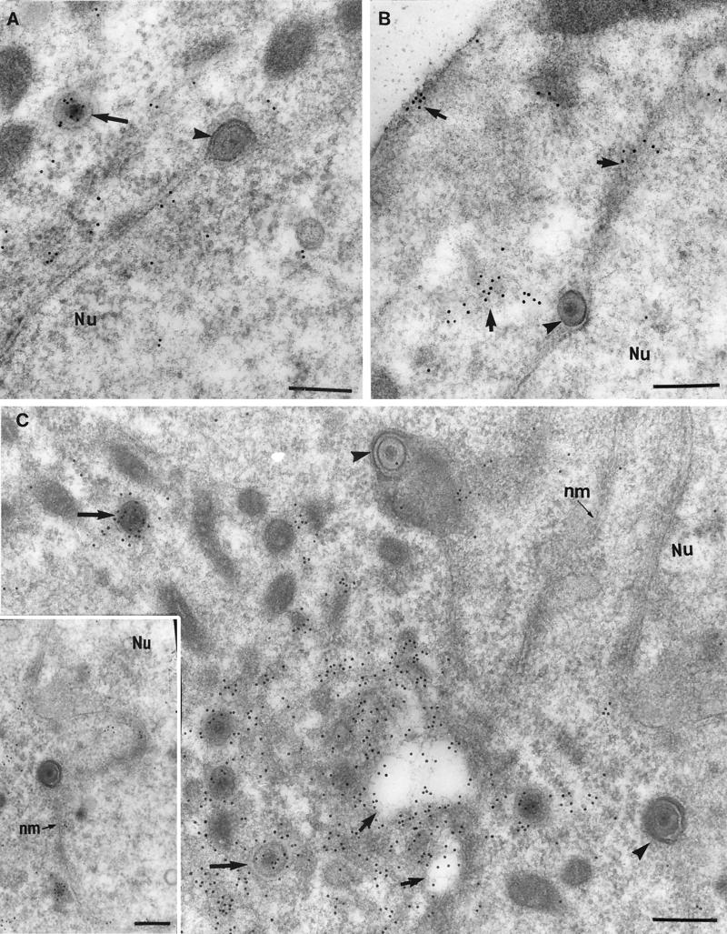
FIG. 3.
Comparison of tegument and glycoprotein labeling of budding and cytoplasmic viral particles (12 hpi). Neurons were embedded in Lowicryl HM20 and reacted with polyclonal antibodies against VP22 (A), gD (B), and US9 (C). (A) Unlabeled budding viral particle (arrowhead) and intracytoplasmic enveloped virion (arrow) densely labeled with VP22. Bar, 200 nm. (B) Plasma membrane, cytoplasm, and nuclear membrane (arrows), but not perinuclear enveloped virions (arrowhead) labeled with gD. Bar, 200 nm. (C) Intracytoplasmic enveloped virions (larger arrows) and vesicular regions (smaller arrows) close to the nucleus (Nu) but not the budding virons (arrowheads) labeled densely with US9. Bar, 200 nm. The inset shows a complete view under lower magnification of the virion (at the bottom right corner in Fig. 3C) budding from the nucleus. Bar, 200 nm.

