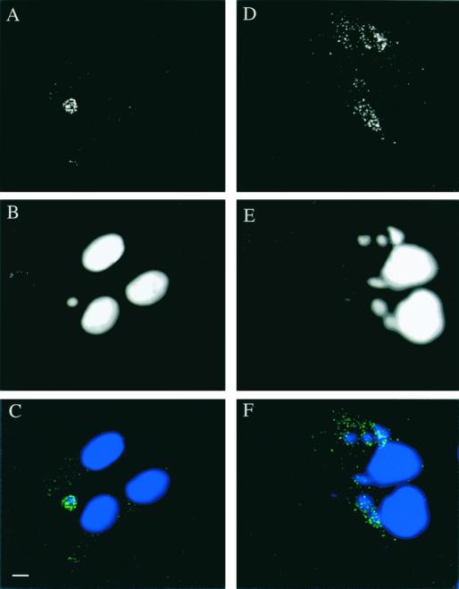
FIG. 4.
Immunofluorescence localization of E8R in infected HeLa cells at 3 h postinfection (A, B, and C) and at 6 h postinfection (D, E, and F). (A and D) Labeling with antibodies to E8R (green channel in C and F). (B and E) Labeling with Hoechst stain (blue channel in C and F). (C and F) Merges. The labeling shows a fluorescence-labeled ring around the viral factories (C). Late in infection, however, the labeling shows a granular pattern that does not colocalize with the viral factories (F). Bar, 2 μm.

